
A systematic review examines cases with Lemierre’s syndrome (LS) in the past 5 years.
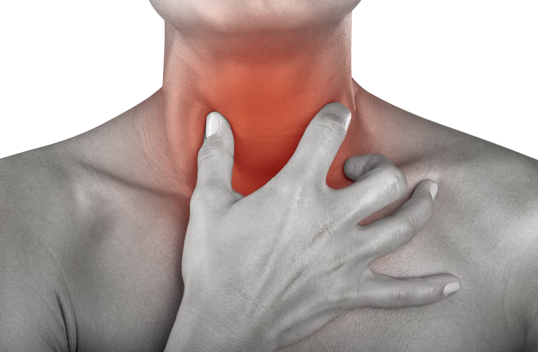
LS is characterized by sepsis often evolving after a sore throat or tonsillitis and then complicated by various septic emboli and thrombosis of the internal jugular vein. Symptoms include sepsis, pain, and/or swelling in the throat or neck, as well as respiratory symptoms.
Laboratory findings show elevated infectious parameters and radiological findings show thrombosis of the internal jugular vein and emboli in the lungs or other organs. The syndrome is often associated with an infection with Fusobacterium necrophorum.
Researchers found a total of 137 cases of LS, of which 47 were infected with F. necrophorum and others with Staphylococcus and Streptococcus. Complications of this rare but severe disease included osteomyelitis, meningitis, and acute respiratory distress syndrome. Mortality was extremely high in the pre-antibiotic era but has diminished with the advent of antibiotics.
The review showed a mortality rate of only 2% of which none of the cases involved fusobacteria. Duration of treatment varied; a 4–6-week course of carbapenem or piperacillin/tazobactam in combination with metronidazole was optimum. Other treatment options included anticoagulants in 46% of cases, which is unwarrantedly high, as to date, no evidence of the positive effects of anticoagulants in LS exists. Only two cases had ligation of the internal jugular vein performed. This review confirms the rare, but severe aspects of LS. Mortality from LS in this day and age appears to be low, however the syndrome is difficult to recognize, and still requires the full attention of the clinician.
Diagnosis
A definite diagnosis of LS should be made based on the following findings:
1) a recent pharyngeal illness
2) complicated by septic emboli
3) as well as either thrombosis of the internal jugular
Common clinical characteristics of LS
- Septic emboli (100%)
- Thrombosis of the internal jugular vein (84%)
- Sepsis (50%)
- A sore throat (24%)
- Swelling of the neck (5%)
- CNS affection (3.6%)
Imaging
Concerning the diagnosis of the infectious thrombosis, the optimal imaging tool is still debated. Ultrasonography is a radiation-free and easily available bedside tool, but it has the disadvantage of being less sensitive for recently formed thrombosis material, with a lesser echogenicity, as well as difficulties in the deeper tissue areas around the clavicle and mandible. MRI is an excellent method for visualizing all anatomic structures as well as the thrombosis and/or septic emboli. However, MRI is expensive and usually not readily available. CT scans are cheaper and available in most hospitals, but it involves radiation exposure.
Management Antibiotics
All but a few rare cases received antibiotic treatment during their hospital stay. Multiple different regimens of the antibiotic of choice were used. Resistance to penicillin was not found at an increased frequency. However, carbapenem and piperacillin/tazobactam were commonly used, either as monotherapy or in combination with metronidazole, and 98% of the cases (mortality n=2) were treated successfully. The mean duration of antibiotic treatment was 4 weeks, but it ranged from 10 days to 8 weeks.
Anticoagulation
Anticoagulation is still a controversial issue, with no proper studies carried out on the issue. In this review, 87 cases (64%) received anticoagulation treatment, often with low-molecular-weight heparin. Treatment duration varied between 2 weeks and 6 months.
Other forms of treatment
A substantial number of cases (n=17) had abscesses in various anatomical locations: lungs, liver, epidura, and neck. Generally, accessible abscesses were treated by drainage. Rarely, other surgical procedures were performed: tooth extraction, craniotomy, and ligation of the occluded vein in order to prevent further septic emboli.
Source: Adapted from: Infection & Drug Resistance