
Together with maxillofacial surgeon, Erik Nout, MD, DDS, PhD, I prepared a stereolithography file of the above 3D-printed mandible.
Speaking about the 3D print, Dr. Nout says:
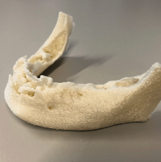
“The 3D-printed mandible is from an 84-year-old patient who presented with exposed mandibular bone in the symphysial area and on the lingual side distally on the right side due to long-term use of bisphosphonate treatment intravenously because of metastasized prostate carcinoma. Remarkably, this patient had no complaints of pain or discomfort whatsoever. Clinically, the gingiva is penetrated and debris is present. Conforming to the guidelines on bisphosphonate treatment, we tried to be as conservative as possible. Because of the abundant amount of debris and the large area of bone exposition, I decided to make a cone beam CT (CBCT) scan. The CBCT scan showed a large sequestration in the symphysial area and a far-reaching mottled aspect of the mandibular bone from region 45-35. It was concluded that the chance of fracture of the mandible was evident. In respect of the age, the absence of complaints, and the medical history of the patient, I decided to make a 3D print of the mandible to prepare ourselves in case of fracture of the mandible. With the acquired 3D model, we were able to bend a reconstruction plate on the mandible in advance to ensure anatomical alignment and re-positioning of the mandible in case of fracture. It also allowed us mimic the anatomical shape of the mandible when we were forced to perform a segmental resection of the mandible. With 3D models, surgery can be better prepared and surgery time is shorter.”
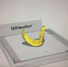 I prepared the file using the Ultimaker 3 with polylactic acid (PLA) and support by polyvinyl alcohol (PVA). It was printed in a ratio of 1:1 and ready in only 4 hours.
 PWeekly