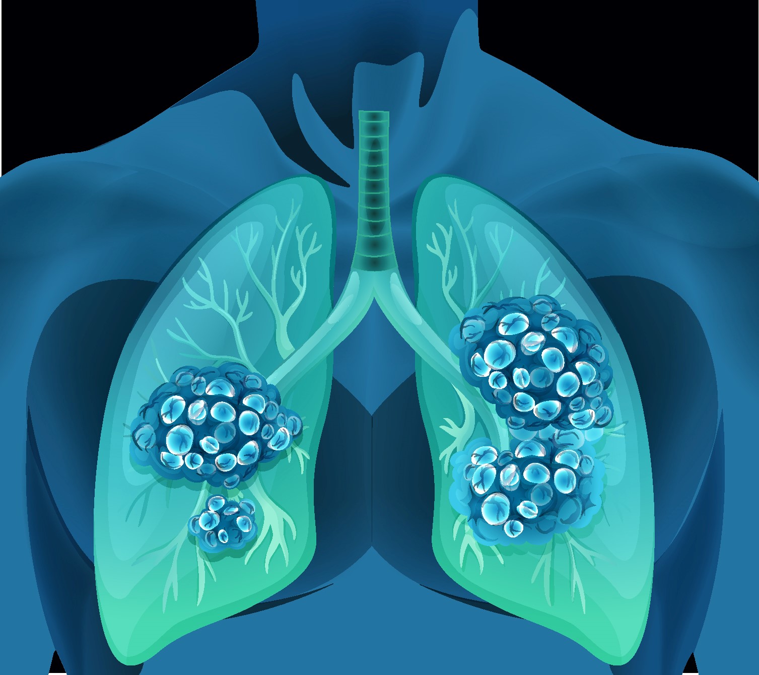
For a study, invasive fungal infections were extremely dangerous and almost always occurred in severely immunocompromised patients, such as solid organ and hematopoietic stem cell transplant recipients, patients with profound and prolonged neutropenia, patients with diabetes mellitus (particularly with ketoacidosis), and others. In these individuals, invasive aspergillosis was the most frequent invasive fungal infection.
However, among other factors, the use of antifungal prophylaxis led to rising in non-Aspergillus invasive fungal diseases, such as mucormycosis. Mucormycosis showed clinically and radiographically similar to invasive aspergillosis, but differentiating between the two diseases was critical since the usual medication for invasive aspergillosis, voriconazole, was ineffective against mucormycosis. Although microbiological identification of the organism was often required for the diagnosis of mucormycosis, several computed tomographic features, such as the reverse halo sign, multiple (>10) nodules, micronodules, and pleural effusion, may favor the diagnosis of mucormycosis over invasive aspergillosis.
When a confirmed tissue diagnosis was not feasible, such differentiation may be highly beneficial. Mucormycosis treatment focused on reversing recognized risk factors, surgical debridement of necrotic tissue, and adequate antifungal medication. Iron chelation treatment may be beneficial in certain people.
Reference:journals.lww.com/clinpulm/Abstract/2016/07000/Cavitary_Lung_Disease_in_a_Heart_Transplant.7.aspx


