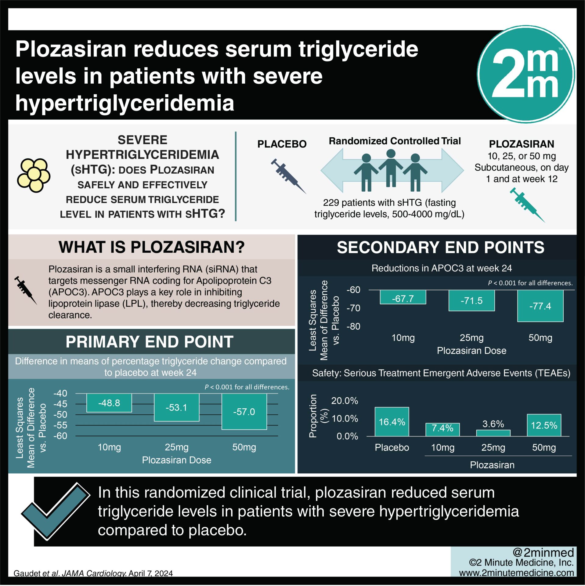
The following is a summary of “Comparison of bone mineral density in forearm and hip regions and evaluation of area of these 2 sites,” published in the December 2022 issue of Primary care by Rajaei, et al.
In middle-aged and older persons, osteoporosis is a frequent condition. Accurate measurement of the surface of the examined region is crucial since bone mineral density (BMD) is calculated by dividing bone mineral content by area. For a study, researchers sought to investigate the hip and forearm areas depending on gender and height.
In cross-sectional descriptive research of 758 participants (702 female and 56 male, separated into 2 groups of ≥50 years and <50 years), skilled workers used a Hologic instrument to perform densitometry on the forearm and femur. SPSS software version 21 was used to evaluate the results statistically.
One-third of the forearm BMD in white women ≥50 years old exhibited moderate agreement with the femoral neck BMD, and overall forearm BMD in this group also showed moderate agreement. One-third of the forearm BMD in Caucasian women < 50 years old showed good agreement with the femoral trochanter. Total forearm BMD in the same sample of people likewise showed extremely strong agreement with the femoral trochanter. Trochanter, intertrochanteric, neck, and total forearm BMD exhibited strong agreement with all 4 areas of the femur in women <50 of white race, while total forearm BMD showed extremely good agreement with all 4 regions of the femur in the same group of people.
It appeared that simultaneous measurement of the one-third forearm region and various hip areas enhanced the accuracy of total BMD measurement, at least based on the findings of the comparison of the forearm one-third with hip areas.
Reference: journals.lww.com/jfmpc/Fulltext/2022/12000/Comparison_of_bone_mineral_density_in_forearm_and.27.aspx