Lid wiper epitheliopathy (LWE) has received more attention during the diagnosis and treatment of the dry eye. However, its causes and pathogenesis remain unclear. We aimed to explore the etiology of LWE by analyzing the association between the severity of LWE and different anatomical and tissue morphological examination characteristics using confocal microscopy on eyes with dry eye syndrome.
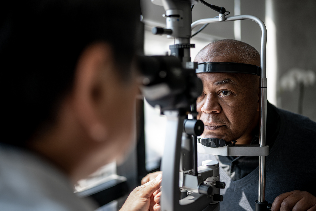
We recruited 350 patients with LWE and dry eye syndrome (350 eyes). We examined the eyes with lid-wiper staining, conjunctival staining, a comprehensive ocular surface exam using the OCULUS keratography 5M, conjunctival impression cytology, and confocal microscopy observations. We analyzed the associations between each indicator and the LWE staining score.
According to the Spearman’s analysis, the LWE staining score was weakly associated with thickness of the lipid layer (r=0.1737, p=0.0005) and severity of Meibomian gland dysfunction (r=0.2026, p<0.0001); and strongly associated with staging of conjunctival impression cytology (r= -0.7694, p<0.0001). Pearson's correlation analysis indicated that, LWE staining score was moderately associated with age (r=0.4165, p<0.0001), tear meniscus height (r=0 -0.4019, p<0.0001), and NIKBUT-first (noninvasive keratography tear film breakup time) (r= -0.5108, p<0.0001); and strongly associated with NIKBUT-average (r= -0.7820, p<0.0001) and ocular staining score (r=0.6113, p<0.00001). Some patients presented abnormal blinking. We observed deeper lesion depths and more holes and fissures in the lid wipers of patients with more severe LWE than in patients with milder LWE.
Abnormal friction factors caused by insufficient lubrication between the lid wiper area and the ocular surface seem to influence the development and/or the severity of LWE. Aggravation of LWE further increases the frictional damage between the lid wiper and the ocular surface.
Analysis of factors leading to lid wiper epitheliopathy.