MONDAY, Aug. 5, 2019 (HealthDay News) — In a scientific expert panel consensus document, published in the July 16 issue of the Journal of the American College of Cardiology, recommendations are presented for how cardiac magnetic resonance (CMR) imaging after myocardial infarction (MI) is used in clinical research.
Borja Ibañez, M.D., Ph.D., from the Centro Nacional de Investigaciones Cardiovasculares in Madrid, and colleagues offer recommendations for CMR end point selection in experimental and clinical trials based on pathophysiology and its association with outcomes.
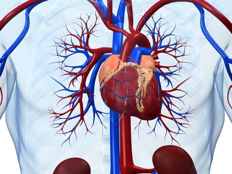
The authors note that CMR has become indispensable for post-MI tissue characterization. It is recommended that the index CMR scan be performed five ± two days after reperfusion. Several key parameters have been shown to be relatively stable at this time point. The extent of late gadolinium enhancement (LGE) is the recommended CMR primary end point, expressed in both absolute (grams) and relative (percentage of left ventricular [LV] mass) terms. The main secondary end points are percentage LV ejection fraction and microvascular obstruction (areas of hypoenhancement within LGE in grams or as percentage of LV).
“Currently, many clinical trials use magnetic resonance imaging to assess a principal outcome, but it is very difficult to compare these studies because they use widely different protocols,” Ibañez said in a statement. “Myocardial infarction affects millions of people in the world every year, and this is therefore a highly active field of research. Because of this, the implications of the new consensus document are enormous.”
Several authors disclosed financial ties to the pharmaceutical and medical device industries.
Copyright © 2019 HealthDay. All rights reserved.