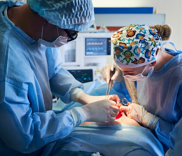
The local and regional radiographic results of MI transforaminal lumbar interbody fusion (TLIF) versus open TLIF remain unknown. A study aimed to thoroughly evaluate local and regional radiography characteristics after MI-TLIF and open TLIF. The authors hypothesized that open TLIF corrects more segmental and global lordosis than MI-TLIF. A retrospective cohort analysis of consecutive patients undergoing MI- or open TLIF for grade I degenerative spondylolisthesis was carried out in a single location. Patients who received open TLIF were matched to those who underwent MI-TLIF using one-to-one nearest-neighbor propensity score matching (PSM). Segmental lordosis (SL), anterior disc height (ADH), posterior disc height (PDH), foraminal height (FH), % spondylolisthesis, and cage position were all measured on a sagittal segmental radiograph. Overall lumbar lordosis (LL), pelvic incidence (PI)–lumbar lordosis (PI-LL) mismatch, sacral slope (SS), and pelvic tilt were lumbar radiography characteristics studied (PT). Following surgery, changes in segmental or total lordosis were called “lordosis” if they were greater than 0° and “kyphosis” if they were less than 0°. To compare outcomes between the MI-TLIF and open-TLIF groups, student t-tests or Wilcoxon rank-sum tests were performed.
A total of 267 patients were included in the study, 114 (43 % ) who received MI-TLIF and 153 (57 % ) who underwent open TLIF, with an average follow-up of 56.6 weeks (SD 23.5 weeks) (SD 23.5 weeks). There were 75 patients in each group after PSM. At the most recent follow-up, both MI- and open-TLIF patients improved significantly on the Oswestry Disability Index (ODI) and the numeric rating scale for low-back pain (NRS-BP), with no significant differences between groups (p > 0.05). Compared to baseline, both MI- and open-TLIF patients had significant improvements in SL, ADH, and % corrected spondylolisthesis (p<0.001). However, the MI-TLIF group saw considerably greater magnitudes of correction in these measures (SL 4.14°± 4.35° vs 1.15° ±3.88°, p<0.001; ADH 4.25± 3.68 vs 1.41± 3.77 mm, p<0.001; % corrected spondylolisthesis: 10.82% ±6.47% vs 5.87% ±8.32 %, p<0.001). LL improved in 44 % (0.3°± 8.5°) of the cases in the MI-TLIF group, compared to 48 % (0.9°± 6.4°) of the cases in the open-TLIF group (p>0.05). Stratification based on operational approach (unilateral vs. bilateral facetectomy) and interbody device (static vs. expandable) produced no statistically significant differences (p>0.05). Patients undergoing MI or open-TLIF exhibited significant improvements in patient-reported outcome (PRO) measures and local radiographic parameters, with little influence on regional alignment. Surprisingly, change in SL was much greater in MI-TLIF patients in the sample, possibly reflecting the effect of operational procedures, technical advancements, and the preservation of the posterior tension band. There were no significant overall changes in LL between groups, implying that MI-TLIF is comparable to open methods in delivering radiographic correction after surgery, taking these findings together. These findings demonstrate that either MI- or open-TLIF methods can accomplish alignment aims, emphasizing the necessity of surgeon attention to these aspects.


