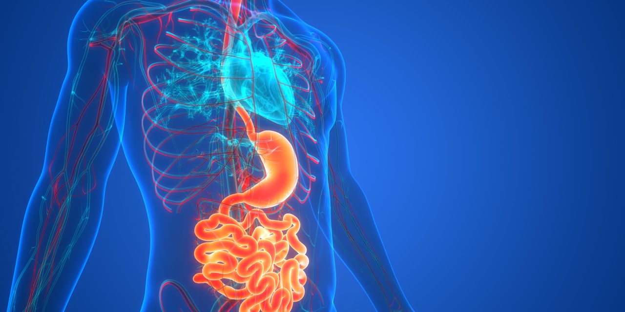
The homeostatic proliferation-differentiation gradient in the esophageal epithelium is perturbed under inflammatory disease conditions such as gastroesophageal reflux disease and eosinophilic esophagitis. Herein we describe the protocols for rapid generation (<14 days) and characterization of single-cell-derived, three-dimensional (3D) esophageal organoids from human subjects and mice with normal esophageal mucosa or inflammatory disease conditions. While 3D organoids recapitulate normal epithelial renewal, proliferation, and differentiation, non-cell autonomous reactive epithelial changes under inflammatory conditions are evaluated in the absence of the inflammatory milieu. Reactive epithelial changes are reconstituted upon exposure to exogenous recombinant cytokines. These changes are modulated pharmacologically or genetically ex vivo. Molecular, structural, and functional changes are characterized by morphology, flow cytometry, biochemistry, and gene expression analyses. Esophageal 3D organoids can be translated for the development of personalized medicine in assessment of individual cytokine sensitivity and molecularly targeted therapeutics in esophagitis patients © 2020 by John Wiley & Sons, Inc. Basic Protocol 1: Generation of esophageal organoids from biopsy or murine esophageal epithelial sheets Basic Protocol 2: Propagation and cryopreservation of esophageal organoids Basic Protocol 3: Harvesting of esophageal organoids for RNA isolation, immunohistochemistry, and evaluation of 3D architecture Basic Protocol 4: Modeling of reactive epithelium in esophageal organoids.© 2020 John Wiley & Sons, Inc.
Modeling Epithelial Homeostasis and Reactive Epithelial Changes in Human and Murine Three-Dimensional Esophageal Organoids.


