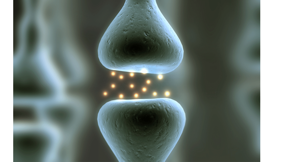
A comparison of two emerging blood markers of neurodegeneration favored neurofilament light (NfL), a marker of axonal injury, over total plasma tau for predicting cognitive decline and imaging changes, a longitudinal study showed.
Although associations with cognition and imaging were more strongly associated with NfL than total tau, baseline plasma total tau strengthened cross-sectional associations, particularly with respect to memory-specific decline, reported Michelle Mielke, PhD, of the Mayo Clinic in Rochester, Minnesota, and colleagues, at the 2021 American Academy of Neurology annual meeting.
Overall, plasma NfL had better utility as a prognostic marker of cognitive decline and neuroimaging changes, suggesting it may help determine how fast someone declines and how effective future therapies might be in slowing this decline.
Plasma T-tau added cross-sectional value to NfL in specific contexts, but longitudinally, it did not add to the prognostic value of NfL. As a diagnostic tool, however, it may be useful to add measurement of total tau to NfL, Mielke noted.
“Understanding which marker is associated with which aspect of neurodegeneration will inform how each marker can best be used for specific clinical purposes and what information each marker provides,” she said in a statement.
The team studied 995 patients enrolled in the Mayo Clinic Study on Aging for a median of 6.2 years, obtaining neuroimaging, cognitive testing, and plasma levels of both markers about every 15 months. Models were adjusted for age, sex, and education.
Cross-sectional associations for the combination of NfL and total tau at baseline included:
- Cognitive measures: global cognition (memory, attention, verbal fluency, language, and the ability to understand images and interpret spatial relationships) and memory-specific scores.
- Imaging: reduced temporal cortex thickness and increased number of infarcts.
Neurodegeneration occurs in many disorders. In the central nervous system, examples include multiple forms of dementia including Alzheimer’s disease, frontotemporal dementia, vascular dementia, and Lewy body dementia. Causes and locations of neurodegeneration in these conditions are variable.
Assessing neurodegeneration is important in the biological definition of Alzheimer’s disease (the National Institute on Aging and Alzheimer’s Association research framework of ATN staging) with implications for diagnosis, prognosis, and developing effective disease-modifying treatments. Means of assessing neurodegeneration include autopsy and, in the living, biopsy, imaging with both MRI and PET scans, and cerebrospinal fluid studies of markers of neuronal injury. Measurable blood markers would be easier to obtain and valuable if they correlated with important clinical variables such as cognitive scores and assessments of neurodegeneration.
During the last decade, NfL and tau have emerged as blood-based neurodegeneration marker candidates with particular relevance in the area of dementia. This was made possible by the development of sensitive assays such as the Simoa platform, extending earlier work with other assay types.
A 2020 study related plasma NfL to PET and MRI measurements of brain atrophy (neurodegeneration). Cross-sectional plasma NfL associations with PET changes in brain regions typically affected by Alzheimer’s disease were specific to amyloid PET in a cognitively unimpaired cohort, but with tau PET in people who were cognitively impaired. “Plasma NfL increases in response to amyloid-related neuronal injury in preclinical stages of Alzheimer’s disease, but is related to tau-mediated neurodegeneration in symptomatic patients,” the authors observed.
Tau is the major microtubule-associated protein in mature neurons and is pathologically implicated in Alzheimer’s disease, frontotemporal dementia, and other disorders. In Alzheimer’s, hyperphosphorylated tau aggregates are the core of neurofibrillary tangles, a pathologic hallmark of the disorder. These tau tangles are more closely associated with cortical atrophy and clinical symptoms than amyloid beta in Alzheimer’s disease.
Several recent studies have considered the combination of NfL and total tau in Alzheimer’s disease. A retrospective 2020 study considered the utility of plasma NfL and total tau for clinical trials in Alzheimer’s disease and emphasized the potential for pre-clinical NfL changes to be useful in trials of disease-modifying therapy. NfL had a stronger association than total tau with clinical scores and did not offer additional information to that given by NfL (P>0.05 at all time points).
A 2020 longitudinal study considering NfL and total tau in Alzheimer’s disease found baseline plasma NfL was higher in Alzheimer’s dementia than both mild cognitive impairment and normal cognition. NfL did not predict conversion to dementia, though it did predict decline on three of 10 neuropsychological tests. Plasma total tau was higher in Alzheimer’s dementia than normal cognition but not mild cognitive impairment, and it predicted no longitudinal outcomes.
“Studies have shown that cross-sectionally and longitudinally, elevated levels of plasma T-tau and NfL are associated with worse cognition and neuroimaging measures of cortical thickness, cortical atrophy, white matter hyperintensity, and white matter integrity,” Mielke and co-authors noted.
In the Mayo study, no associations were seen for the combined markers of NfL and tau with hippocampal volume, white matter integrity, and white matter hyperintensity volume.
Mielke and colleagues group replicated their analysis of Mayo Clinic participants in a cohort of 387 patients without dementia from the multicenter Alzheimer’s Disease Neuroimaging Initiative (ADNI) cohort who were followed for a median of three years, with similar outcomes, suggesting the findings may be replicated across clinical settings.
-
A comparison of two emerging blood markers of neurodegeneration favored neurofilament light (NfL), a marker of axonal injury, over total plasma tau for predicting cognitive decline and imaging changes, a longitudinal study showed.
-
Assessing neurodegeneration is important in the biological definition of Alzheimer’s disease with implications for diagnosis, prognosis, and developing effective disease-modifying treatments.
Paul Smyth, MD, Contributing Writer, BreakingMED™
This research was funded by National Institutes of Health and National Institute on Aging grants, the GHR Foundation, and the Rochester Epidemiology Project.
Mielke reported consulting relationships with Brain Protection Company and with Biogen.
Cat ID: 131
Topic ID: 82,131,282,730,131,33,361,192,255,925,167


