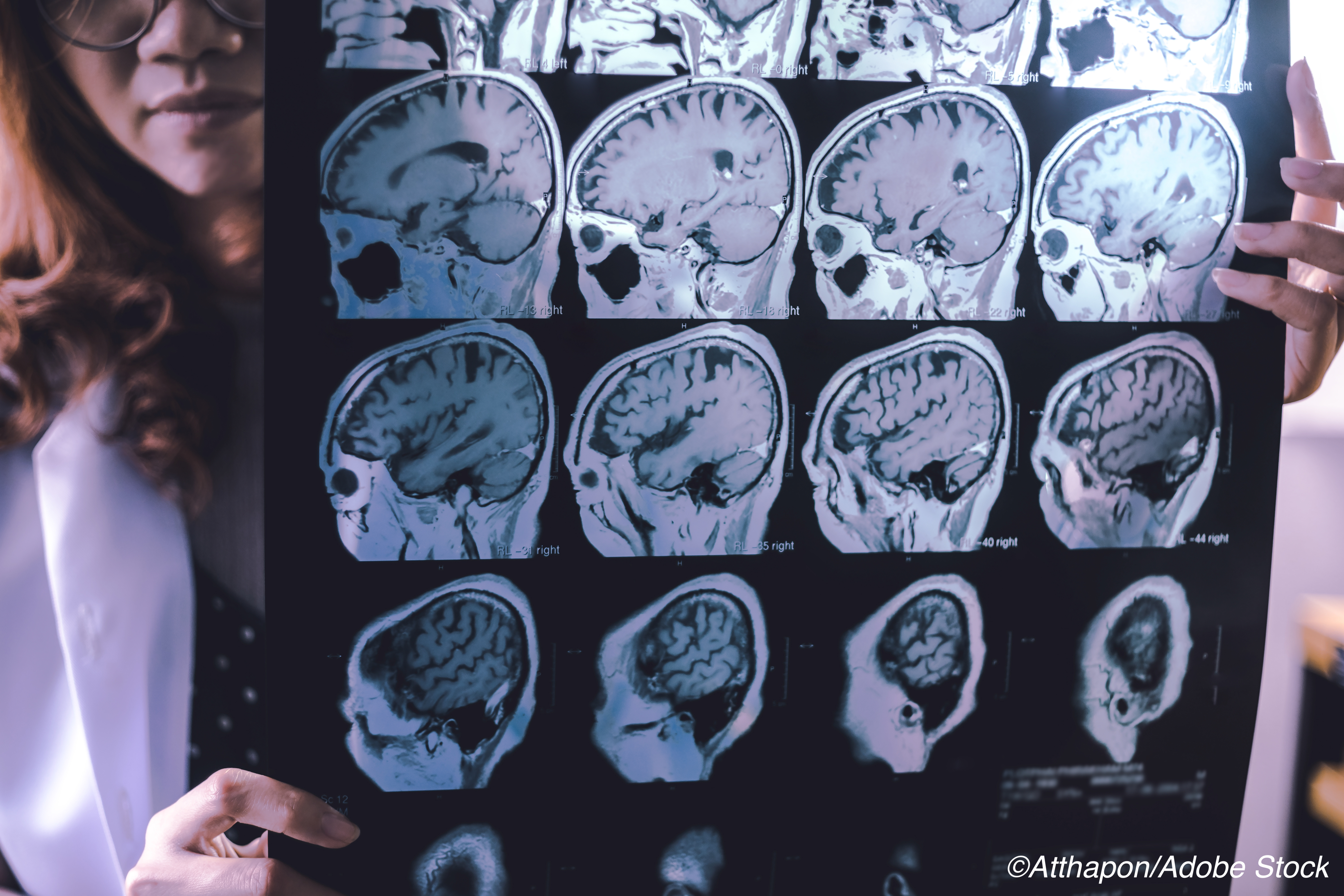
Intranasal oxytocin, with or without instructed facial mimicry, increased functional MRI (fMRI) signals in brain areas associated with emotional processing and empathy in patients with frontotemporal dementia (FTD), a double-blind, randomized, placebo-controlled crossover trial found.
“Oxytocin and imitation, alone or in combination, activate frontal and other limbic brain regions in patients with FTD,” wrote Elizabeth Finger, MD, of Western University in London, Ontario, and coauthors in Neurology.
“Importantly, this provides novel evidence that latent capacity is present, even in patients with significant neurodegeneration, in brain regions affected early in the disease course,” they added.
Increased blood-oxygen-level dependent (BOLD) activity when viewing or imitating facial expressions was observed after oxytocin versus placebo in frontal and limbic regions including bilateral anterior insular, bilateral inferior frontal gyrus, bilateral caudate, right anterior cingulate cortex, and right inferior parietal lobule.
Combination treatment with oxytocin and instructed mimicry was associated with increased responses in these regions and in the right amygdala. Instructed mimicry alone showed greater activation in bilateral inferior frontal gyrus and inferior parietal lobule, for both FTD patients and controls.
“The present results warrant further investigation of oxytocin, as well as other pharmacologic and behavioral approaches, to augment empathy and related social cognitive processing to improve key symptoms of FTD,” wrote Hitoshi Shinotoh, MD, PhD, of the National Institutes for Quantum and Radiological Science and Technology, in Chiba, Japan, in an accompanying editorial.
“An ongoing phase II randomized clinical trial of oxytocin in patients with FTD will provide further data regarding whether and how repeated dosing of oxytocin modulates empathy, expression recognition, and related social behaviors,” Shinotoh added.
Abnormal processing of emotional facial expressions is considered a key factor in the empathy deficits observed in FTD, and oxytocin has been proposed as a mediator of social behavior and potential treatment for aberrant social cognition. In a 2011 study of 20 FTD patients, a 24 IU intranasal dose of oxytocin was associated with reduced recognition of angry facial expressions and better caregiver ratings of social behaviors, and a 2015 study of intranasal doses of 24, 48, and 72 IU found Class I evidence that oxytocin was safe and well tolerated in FTD patients.
Facial mimicry, whether spontaneous or instructed, has been considered a means of eliciting empathy, and shown to activate a brain network including the inferior frontal gyrus, insula, and amygdala, with more activity during imitation, compared with observation.
In this research, Finger and colleagues studied 28 patients with FTD identified between 2013 and 2017 (about 54% male, mean age about 64), along with 23 healthy volunteers (about 48% male, mean age about 61), excluding people with stroke or a non-FTD neurologic disorder.
FTD patients had two study visits 2 weeks apart with fMRI and other assessment; those who received placebo (oxytocin) at the first visit received oxytocin (placebo) at the second in the crossover design. Controls had one visit with fMRI and other assessment and were all treated with placebo (though they were informed of this after evaluation).
At each visit, following the last of three intranasal sprays (placebo or a total of 72 IU of oxytocin), participants had fMRI with an in-scanner view-and-imitate task that included facial expressions and hand movements. This was followed by behavioral evaluations outside the scanner that included a measure of empathy and a postural knowledge test (choose the correct gesture to complete a cartoon showing an incomplete action) thought to assess the simulation (mirror neuron) network. The view-and-imitate task was repeated outside the scanner using only six facial expressions.
Comparing oxytocin versus placebo within the FTD group, those receiving oxytocin showed greater accuracy identifying happy versus other facial expressions on the view-and-imitate task, with a modestly higher rating on the empathy test (P=0.013), and greater accuracy on postural knowledge testing.
“While fMRI changes induced by oxytocin and imitation in this cohort were significant and in line with models of enhancing facial expression and empathy-related processing, mimicry or a single dose of 72 IU of oxytocin did not significantly improve expression labeling or self report of empathic feelings when viewing emotional pictures,” the authors noted.
The lack of a measurable benefit on expression recognition accuracy “indicates that although fMRI changes were robust and serve as an objective index of oxytocin effects, additional ecological assessments, particularly of daily behaviors, are needed to determine whether these neural signals will translate into improved symptoms and behavior in real-world situations,” they added.
Limitations include reduced numbers of FTD patients used in some analysis due to inability to complete tests. Also, clinical and radiographic heterogeneity of the FTD cohort may have affected BOLD patterns.
-
Intranasal oxytocin increased functional MRI signals in brain areas associated with emotional processing and empathy in patients with frontotemporal dementia (FTD), a double-blind, randomized, placebo-controlled crossover design trial found.
-
Further assessments are needed to determine whether these neural signals translate into improvements in real-world situations; a phase II trial is underway.
Paul Smyth, MD, Contributing Writer, BreakingMED™
This research was supported by funding from the Canadian Institutes of Health Research, the Ministry of Research and Innovation of Ontario, and CFREF BrainsCAN.
The researchers reported no disclosures. The editorialist reported no disclosures.
Cat ID: 361
Topic ID: 82,361,282,404,361,255,925


