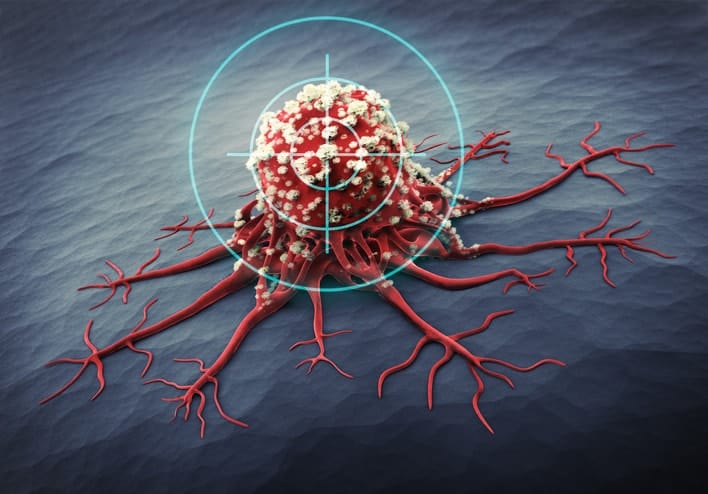
Fusion imaging is gaining attention as an imaging technique to assist minimally invasive tumour ablation. Ultrasound (US) and computed tomography (CT) are the most common imaging modalities to guide thermal ablation of renal tumours, yet cone-beam CT (CBCT) has recently been described to successfully assist percutaneous renal interventions. Our goal was to evaluate primary technical success and correct lesion targeting of US/CBCT fusion imaging to guide the ablation of kidney masses < 2 cm in a small group of patients.
Six renal lesions (maximum diameter 11-17 mm) were treated with RFA in 5 different patients using real-time US/CBCT. Fusion imaging was used to identify and monitor tumour ablation. Demographics, tumour characteristics and mean serum creatinine levels were recorded before and after the procedure. Primary technical success and correct lesion targeting represented the main endpoints of the study. Primary technique efficacy was confirmed at 1-month and 3-month contrast-enhanced CT follow-ups.
In all cases, a confident US/CBCT synchronisation was reached and allowed for a correct targeting and a successful percutaneous ablation. Primary technique efficacy was 100%. No recurrence was observed at the follow-up that ranged from 8 to 26 months (mean 16 months).
US/CBCT fusion proved to be a viable method to precisely guide safe and effective percutaneous thermal ablation in patients with small renal tumours, especially when hardly detectable on US.
• US/CBCT fusion imaging for renal ablation is safe and feasible. • US/CBCT fusion imaging allows for an improved targeting and complete ablation of small RCC with poor US-conspicuity.
Real-time US/cone-beam CT fusion imaging for percutaneous ablation of small renal tumours: a technical note.