The following is a summary of “Lentigo maligna melanoma mapping using reflectance confocal microscopy correlates with staged excision: A prospective study,” published in the FEBRUARY 2023 issue of Dermatology by Dechent, et al.
Incomplete excision and possible recurrence might result from lentigo maligna/lentigo maligna melanoma (LM/LMM) when it presents with subclinical extension, which can be challenging to diagnose preoperatively. The LM/LMM boundaries have been evaluated in preliminary experiments using reflectance confocal microscopy (RCM). For a study, researchers sought to assess the relationship between the LM/LMM subclinical extension determined by RCM and the gold standard histopathology.
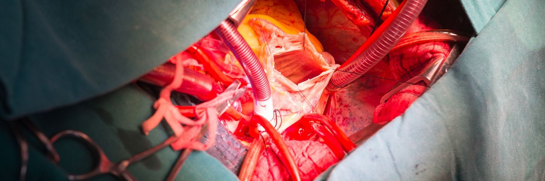
It was prospective research on LM/LMM patients referred for dermatological surgery. Histopathology and RCM results were correlated after a margin-controlled stepwise excision with paraffin-embedded tissue was conducted at the clinically determined initial surgical margin.
The included patients numbered 72. The mean age was 66.8 years (standard deviation, 11.1; range, 38-89); 69.4% of the population was male. 70 of 72 (97.2%), were in the head and neck, with a mean clinical diameter of 1.3 cm (range, 0.3-5). Compared to histology, diagnostic accuracy had a sensitivity of 96.7% and a specificity of 66.7% for detecting residual melanoma in the tumor debulk (after biopsy). An evaluation of the RCM margins showed an overall agreement with the final histopathology of 85.9% (κ = 0.71; P < .001).
The histology of the stepwise excision margins and RCM imaging of the LM/LMM indicated moderate to the excellent overall agreement.
Reference: jaad.org/article/S0190-9622(19)33150-0/fulltext