

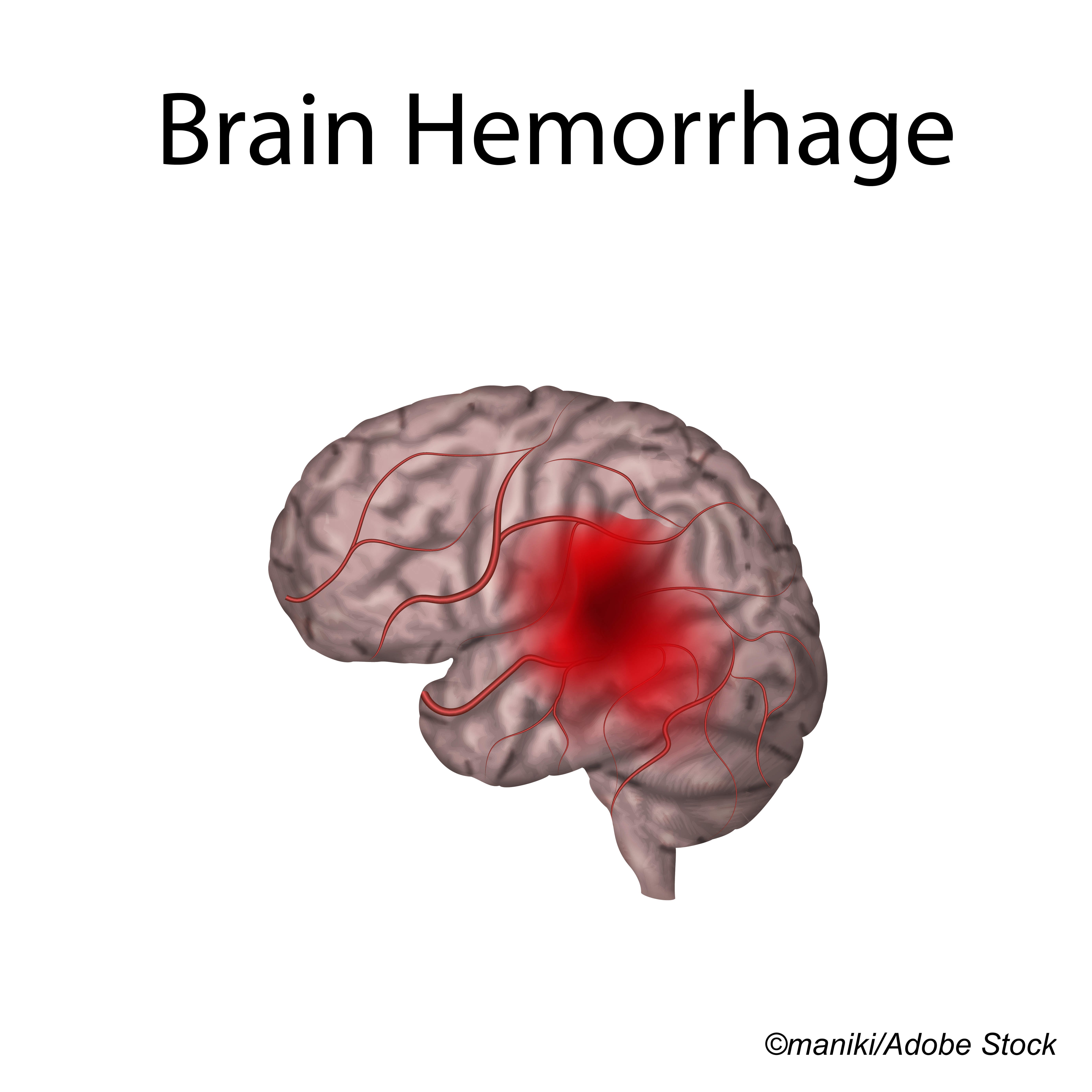
An expert panel consensus statement outlined three recommendations for primary outcomes in pivotal trials of hemostatic agents for acute intracranial hemorrhage.
The optimal primary endpoint was a score on a global patient-centered clinical outcome scale measured between 30 and 180 days after the event, reported Stephan Mayer, MD, of New York Medical College in Valhalla, and co-authors, in JAMA Network Open.
Clinical trials of hemostatic therapy in intracranial hemorrhage ideally should demonstrate improved clinical outcome as well as hemostasis, the panelists noted.
“Standardization of outcome measures in studies of intracranial bleeding and hemostatic therapy will support comparative effectiveness research and meta-analysis, with the goal of accelerating the translation of research into clinical practice,” they wrote. “The three outcome measures proposed in this consensus statement could help this process.”
“In general, end points that involve a patient-centered outcome are superior to pure radiographic outcomes,” they added. “That said, the goal of a hemostatic therapy is to reduce bleeding, and neuroimaging can accurately measure the success or failure of this goal. For this reason, many (but not all) of our authors consider imaging-based measurements of bleeding to be a valid primary end point; all consider this a reasonable biomarker for phase II studies.”
For phase II and phase III endpoints, the group’s recommendations were:
- Best option: A global patient-centered clinical outcome scale at 30 to 180 days, such as the modified Rankin Scale, its utility-weighted variant, or the extended Glasgow outcome scale; “any similar relevant patient-centered outcome measure would be appropriate,” they noted.
- Second option: a combined clinical and radiographic endpoint linking hemorrhage expansion with a poor outcome. One approach might be hemorrhage expansion at 24 hours with accompanying symptoms (e.g., a 4-point worsening on the National Institutes of Health Stroke Scale or meaningful change on any clinically relevant, valid, and reproducible scale). A second approach might be hemorrhage expansion at 24 hours combined with a poor outcome at 30 days or later.
- Third option: a radiographic measure of hemorrhage expansion at 24 hours only. If a dichotomous measure is used with a threshold, the group specified ranges for volume expansion in absolute (6.0 mL-12.5 mL) and relative (20%-33%) terms. “Regardless of any dichotomized end point for hemorrhage expansion, in clinical trials statistical comparison of a continuous absolute or percentage change measurement should always be performed as either a primary or secondary end point because arbitrary dichotomized end points result in loss of information,” they wrote.
“Active bleeding in the emergency department is common when intracranial hemorrhage patients present acutely, especially when they are on anticoagulants,” Mayer noted in an email to BreakingMED.
“Our goal was to support CNS hemostasis research by identifying best practices for trial design and clinical endpoints,” Mayer continued. “Although patient-centered outcomes are always the gold standard, CT allows us to measure hemorrhage volume growth with incredible precision. Our recommendations also support the use of imaging coupled with appropriate clinical endpoints as valid and useful for drug development.”
In their consensus statement, the panelists also provided guidance on taxonomy and terminology. They defined four groups involving spontaneous or traumatic bleeding, each specified as coagulopathic or non-coagulopathic. They distinguished “intracranial hemorrhage” as any bleeding in the intracranial vault and “ICH” as a specific subtype of spontaneous cerebral bleeding confined to the brain parenchyma or ventricles and usually due to hypertension.
“Because the tempo and pathobiology of traumatic and spontaneous bleeding into the brain is different and because anticoagulation can dramatically affect the risk, magnitude, and mechanism of bleeding, we recommend that hemostatic therapies be assessed for the four subgroups separately when possible,” the authors recommended.
“Combining categories may be appropriate and justifiable when evaluating reversal agents for specific forms of anticoagulation, such as vitamin K antagonists and antiplatelet, antithrombin, fibrinolytic, or anti–factor Xa agents,” they added.
Measuring hemorrhage volume is important since hematoma growth correlates with mortality and functional outcome. Typical research strategies have obtained baseline and followup scans at <3 hours (when risk for hematoma expansion is greatest) and at 24 hours. “When measured accurately, this radiographic outcome (or biomarker) should be considered an accurate measure of clinically relevant hemostasis,” the authors wrote.
Trials that compare changes in CNS hemorrhage volume as a primary or secondary end point should adjust for onset-to-CT time and baseline volume, since both variables affect the risk and extent of further bleeding, the authors recommended. For pediatric patients, measuring hemorrhage volume as a percentage of total brain volume standardizes the measurement for different ages and helps account for changing brain volume with age.
The panelists considered manual computerized planimetric measurement as the reference standard for measuring intracranial hemorrhage volume. This technique outlines and determines the area of bleeding present on each CT slice, then multiplies by the number of slices affected. The ABC/2 technique “may be sufficient for use in observational cohort studies and can be very useful for screening patients for eligibility in trials, but should not be used as an end point for clinical trials,” they concluded.
The group’s literature review also pointed to multiple other factors that affect outcome and can vary from study to study based on population and research focus. Investigators were encouraged to adjust analyses for baseline volume and level of consciousness, age, comorbidities, CT-to-treatment time, use of antiplatelet or antithrombotic agents, hemorrhage location, presence of intraventricular hemorrhage, presence of spot sign, or other variables as needed.
Limitations of the consensus statement included its lack of a formal systematic review and evidence quality grading. The panel was split about whether a purely radiographic endpoint was suitable for phase III trials, though all found it acceptable for drug development and phase II studies.
The guidelines do not apply to intra-operative or perioperative neurosurgical bleeding, aneurysmal subarachnoid hemorrhage, or re-rupture, the authors noted. “These specific situations differ substantially from hemorrhage associated with spontaneous ICH or traumatic intracranial hemorrhage,” they wrote.
-
An expert panel consensus statement outlined three recommendations for primary outcomes in pivotal trials of hemostatic agents for acute intracranial hemorrhage.
-
The optimal primary endpoint was a score on a global patient-centered clinical outcome scale measured between 30 and 180 days after the event.
Paul Smyth, MD, Contributing Writer, BreakingMED™
Funding for the workshop was provided by the National Heart, Lung, and Blood Institute and the U.S. Department of Defense.
Mayer reported serving as an independent safety monitor for the National Institute of Neurological Disorders and Stroke FASTEST trial outside the submitted work.
Cat ID: 130
Topic ID: 82,130,501,521,728,730,130,38,192,925