
by Skeptical Scalpel, Physician’s Weekly Top Blogger
One patient, two complications
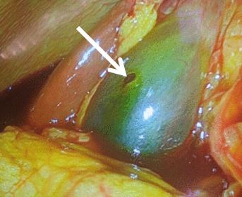
Fig 1. Hole in gallbladder wall
A patient who was to undergo a robot-assisted right nephrectomy because of recurrent pyelonephritis suffered two seldom-seen complications. A Veress needle was placed in the right upper quadrant and bile-tinged fluid was aspirated. The needle was removed and reinserted. During insufflation of carbon dioxide, the patient became unstable. An echocardiogram showed a CO2 embolism.
The patient was immediately placed on his left side in Trendelenburg position and successfully resuscitated. Three days later, the patient developed peritonitis and was taken back to the operating room for laparoscopy. Bile was present in the peritoneal cavity. An inflamed gallbladder was found. After dissection of adhesions, a puncture site in the gallbladder wall was identified. See figure 1.
A laparoscopic cholecystectomy was performed and the patient did well. A nephrostomy tube was later placed to treat the pyelonephritis during recovery from the cholecystectomy. The case report appeared in the journal Trauma Surgery and Acute Care Open.
The authors entitled the paper “Iatrogenic gallbladder perforation secondary to Veress needle placement: A complication of robotic nephrectomy.” I am not a big fan of robotic surgery, but I would not blame the robot for this pair of rare complications.
Use your head
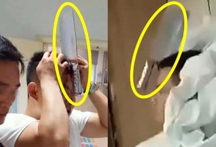
Figure 2. Meat cleaver in head.
A man with a meat cleaver embedded in his head strolled into an emergency department in China. See figure 2. He had been in a fight which presumably he lost. He underwent surgery to remove the knife and was recovering without complications.
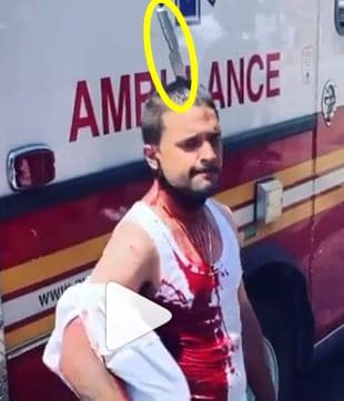
Figure 3. Knife in top of head.
A similar case occurred in The Bronx, New York where a man was stabbed in the head. EMS found him covered in blood and walking around at the crime scene. See figure 3. The events leading to the incident were unclear at the time the article appeared in the New York Post. After surgery to remove the knife, the man was in stable condition at a hospital.
Another recent incident in China involved a man who accidentally shot himself in the chest with a crossbow. As you can see in figure 4, the arrow just missed his heart. Surgery to remove the arrow was successful and he was recovering in a hospital.
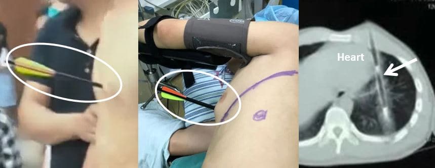
Figure 4. Arrow in left chest. CT scan shows arrow very close to heart.
Be careful walking in your home
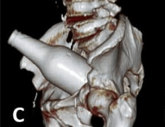
A 67-year-old man presented to a hospital in South America with abdominal pain. He said while walking naked in his house, he lost his balance and fell on a beer bottle that was upright on the floor. A plain x-ray showed a bottle with an air fluid level in the sigmoid colon. A CT scan was done. The reconstructions can be seen in the figures. Plain x-rays of a bottle in the rectosigmoid are not rare, but I’ve never seen a 3-D reconstruction of a bottle before. See Figure 5.
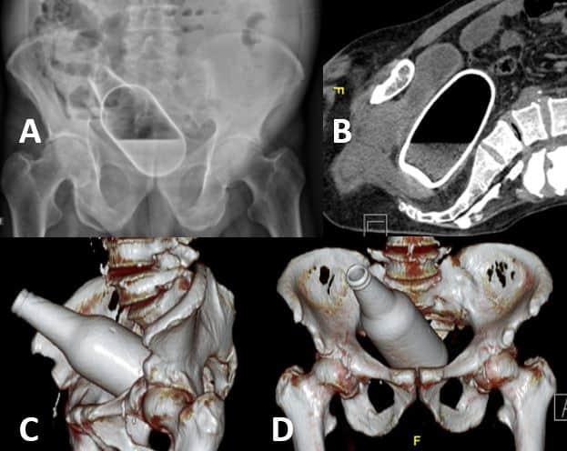
Figure 5. (A) plain x-ray of bottle. (B) CT scan showing bottle in rectosigmoid. (C&D) 3-D reconstructions showing exact position of bottle.
Surgery reveled a hematoma in the lower rectum and a moderate pneumoperitoneum compatible with hollow viscus perforation. A Hartmann procedure was performed.
This case was sent to me on twitter via surgeon @jorgeateo. When I asked him about the history, he replied, “There are no borders; it is always the same answer.”
Related articles
Three unusual cases
Unusual cases part 2
Unusual Cases part 3
Unusual Cases part4
Unusual Cases part 5
The cleaver and crossbow stories were published in The Irish Sun via AsiaWire.
Skeptical Scalpel is a retired surgeon and was a surgical department chair and residency program director for many years. He is board-certified in general surgery and a surgical sub-specialty and has re-certified in both several times.For the last 9 years, he has been blogging at SkepticalScalpel.blogspot.com and tweeting as @SkepticScalpel. His blog has had more than 3,700,000 page views, and he has over 21,000 followers on Twitter.
 SkepticalScalpel