The purposes of this study were to assess the reproducibility of a novel standardized technique for capturing corneal subbasal nerve plexus images with in vivo corneal confocal microscopy and to compare nerve metrics captured with this method in participants with dry eye and control participants.
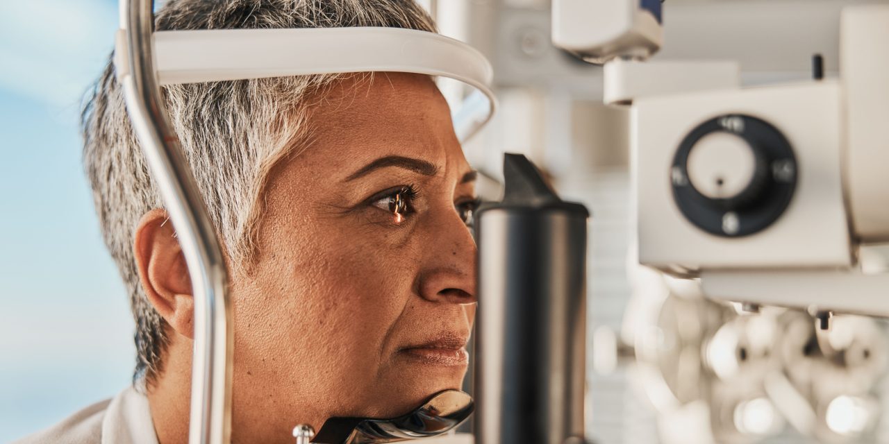
Cases and controls were recruited based on their International Statistical Classification of Diseases and Related Health Problems (ICD-10) diagnoses. Participants completed the following 3 ocular symptom questionnaires: the Ocular Surface Disease Index, Neuropathic Pain Symptom Inventory, and Dry Eye Questionnaire 5. A novel eye fixation-grid system was used to capture 30 standardized confocal microscopy images of the central cornea. Each participant was imaged twice by different operators. Seven quantitative nerve metrics were analyzed using automated software (ACCmetrics, Manchester, United Kingdom) for all 30 images and a 6-image subset.
Forty-seven participants were recruited (25 classified as dry eye and 22 controls). The most reproducible nerve metrics were corneal nerve fiber length [intraclass correlation (ICC) = 0.86], corneal nerve fiber area (ICC = 0.86), and fractal dimension (ICC = 0.90). Although differences were not statistically significant, all mean nerve metrics were lower in those with dry eye compared with controls. Questionnaire scores did not significantly correlate with nerve metrics. Reproducibility of nerve metrics was similar when comparing the entire 30-image montage to a central 6-image subset.
A standardized confocal imaging technique coupled with quantitative assessment of corneal nerves produced reproducible corneal nerve metrics even with different operators. No statistically significant differences in in vivo corneal confocal microscopy nerve metrics were observed between participants with dry eye and control participants.
Validation of a Novel Confocal Microscopy Imaging Protocol With Assessment of Reproducibility and Comparison of Nerve Metrics in Dry Eye Disease Compared With Controls.