
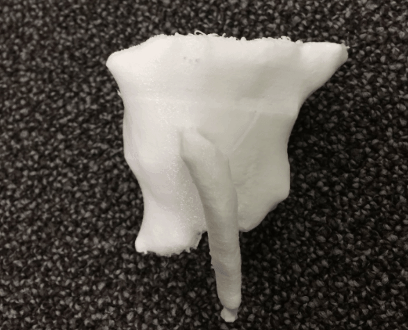
Together with colorectal surgeon David Zimmerman, MD, PhD, FEBS, I’m exploring the world of fistula surgery. We just printed our first and second fistula to investigate the benefits of this type of 3D printing.
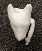
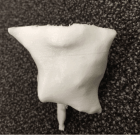
The print process is especially difficult because MRI scans are harder to convert into STereoLithography, or STL, files–which are usually generated by a computer-aided design (CAD) program as an end product of the 3D modeling process–when compared with CT scans. “The main challenge in fistula surgery is to select patients in whom simple treatment (ie, fistulotomy) is feasible,” Explains Dr. Zimmerman. “MRI is usually used to determine patients in whom this is safe without compromise. However, MRI is hard to interpret even for experienced surgeons and radiologists. This case illustrates that 3D printing identifies a fistula that cannot be safely layed open at a simple glance. Also, explaining this difference to patients is much easier when they are able to see their own sphincter and the culprit in one model!”
 PWeekly