
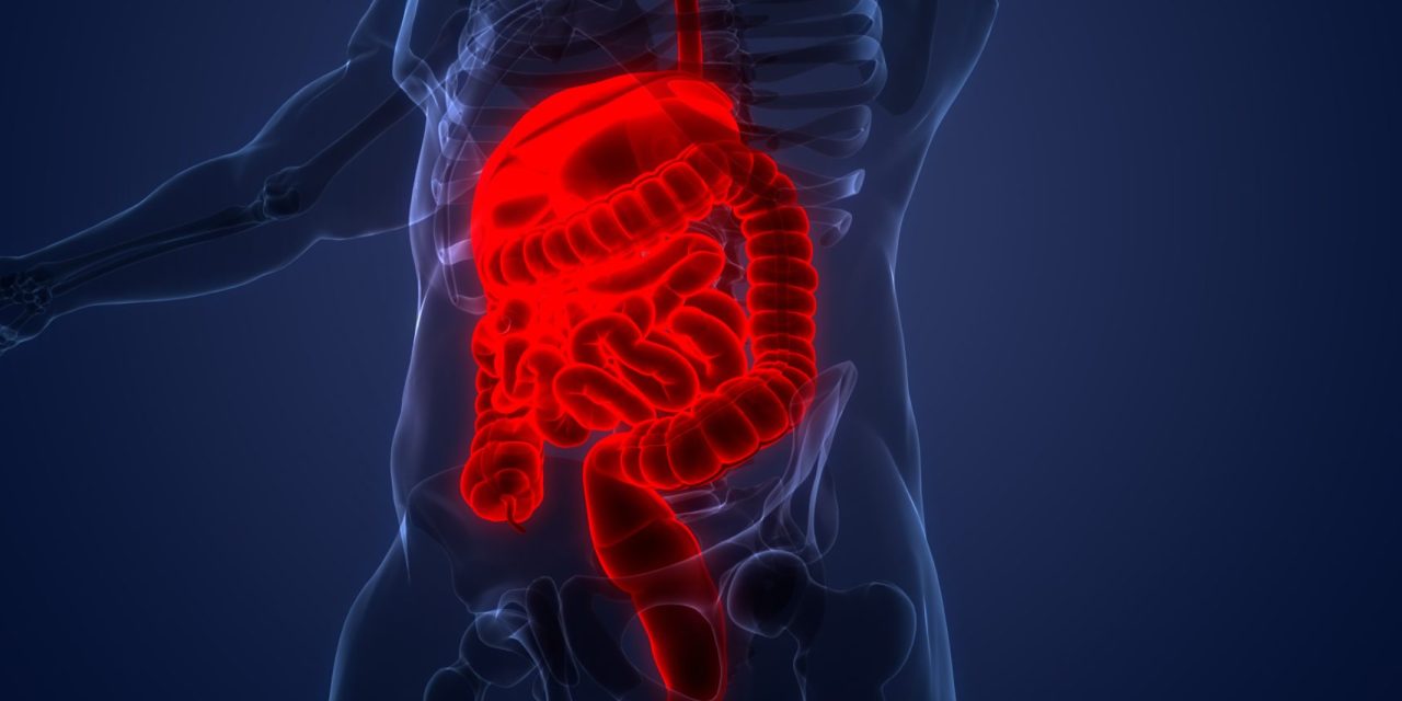
For a study, it was determined that Candida albicans (C. Albicans) was an opportunistic pathogen that caused infections ranging from superficial to life-threatening. C. Albicans could translocate past the gut barrier in a susceptible host, allowing it to spread to deeper organs. Epithelial-driven endocytosis and C. Albicans-driven active penetration were two well-documented methods by which C. Albicans hyphae could have entered human epithelial cells. Autophagy was a method through which host cells protected themselves from intracellular C. Albicans. In myeloid cells, the protective role of autophagy during C. Albicans infection was researched; however, less was known about the role of autophagy during infection of epithelial cells. The involvement of autophagy-related proteins during C. Albicans infection of epithelial cells, including intestinal epithelial cells and gut explants, was determined in the outline. Researchers showed that major autophagy machinery players (LC3-II, PI3P, ATG16L1, and WIPI2) were recruited at Candida invasion sites using cell imaging. Electron microscopy investigations revealed the existence of autophagosomes in the vicinity of invading hyphae, confirming the researcher’s data. Importantly, the activities took place during active C. Albicans penetration into host cells was linked to plasma membrane destruction. Researchers showed that the autophagy-related critical proteins ATG5 and ATG16L1 participated in plasma membrane repair mediated by lysosomal exocytosis and protected epithelial cells against C. Albicans-induced cell death in this environment. The outcomes revealed a novel way by which epithelial cells, which served as the gut’s first line of defense against Candida albicans, might have responded to prevent C. Albicans invasion.
Link:www.tandfonline.com/doi/full/10.1080/19490976.2021.2004798