
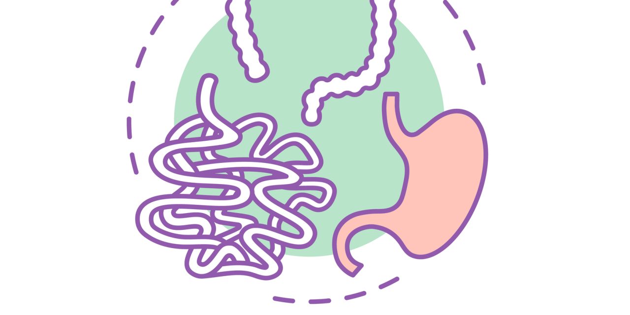
Early detection of children with cystic fibrosis (CF) who are at risk of severe liver disease (CFLD) would allow more focused research into preventative medicines. For CFLD, there is no gold standard test. Ultrasonography (US) is used to diagnose CFLD; however, its diagnostic accuracy is questioned. For a study, researchers sought to determine if variations in routine blood tests, imaging variables, and noninvasive liver fibrosis markers correspond with hepatic US patterns, and if so, how. Researchers looked at baseline research abdominal ultrasound and bloodwork from 244 children with pancreatic insufficiency CF, aged 3 to 12, who were part of a prospective trial to see if ultrasound might predict cirrhosis in CF patients (PUSH study). Children with a heterogeneous (HTG) liver pattern in the US (n=62) were matched 1: 2 in design with children with a normal (NL, n=122) liver pattern in the US. The study of children with nodular (NOD, n=22) and homogeneous hyperechoic (HMG, n=38) livers were studied.
Univariate analysis revealed substantial differences between US groups in routine blood tests, spleen size, and noninvasive liver fibrosis markers. Multivariable models distinguished NOD from NL (AUROC 0.96). Models also discriminate HTG from NL (AUROC 0.76), NOD vs HTG (0.78), and HMG versus NL (0.78). (0.79).
Hepatic US patterns correlated with platelet count, spleen size, and liver fibrosis indices in children with cystic fibrosis. These biomarkers’ multivariable models have the great discriminating capacity for NL versus NOD and decent ability to discriminate other US patterns, suggesting that US patterns correspond with clinically significant liver disease.
Reference:journals.lww.com/jpgn/Fulltext/2019/09000/Liver_Ultrasound_Patterns_in_Children_With_Cystic.19.aspx