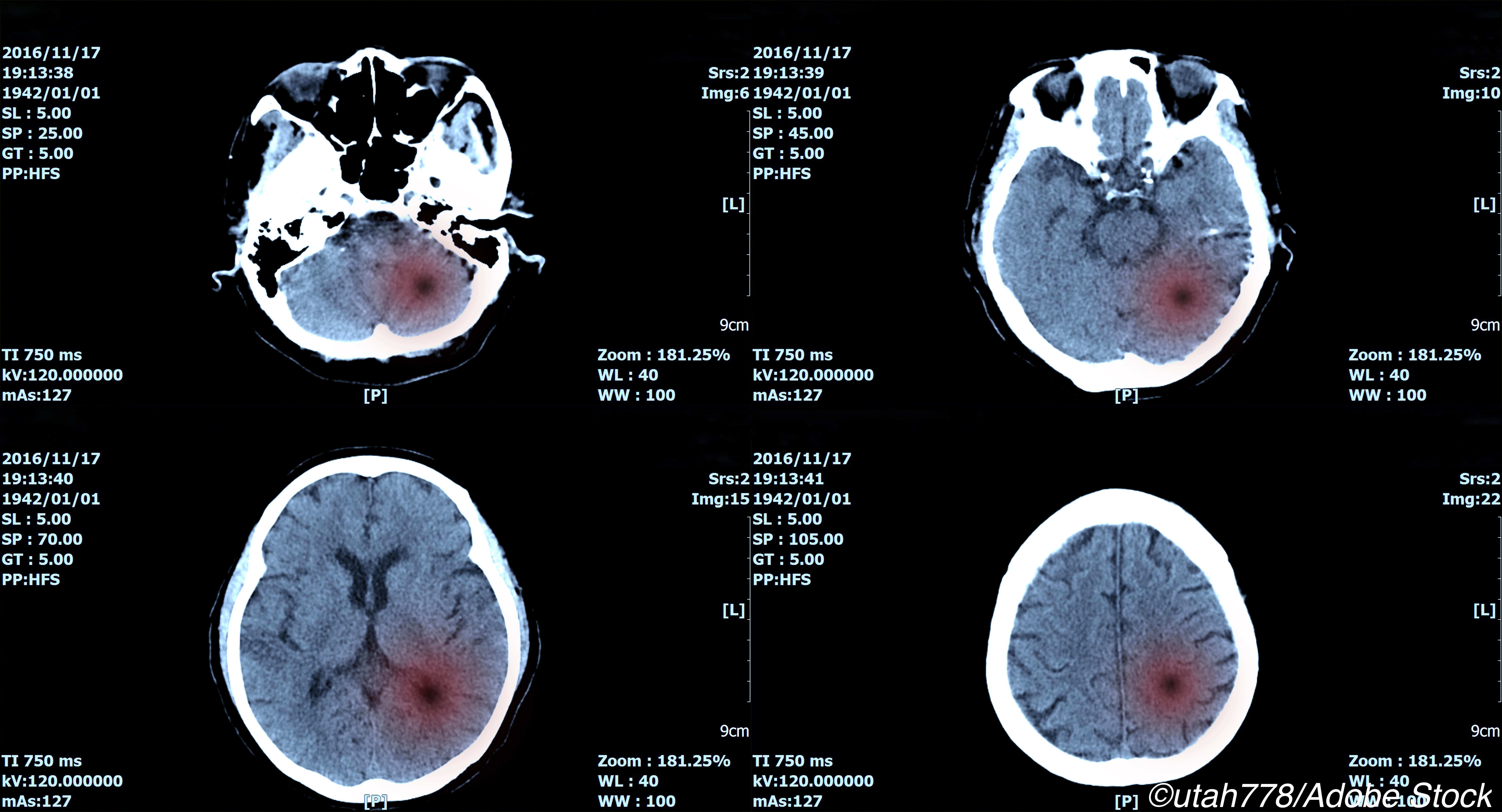
Multiple non-contrast CT (NCCT) markers in intracerebral hemorrhage (ICH) were associated with hematoma expansion and functional outcomes, a meta-analysis found.
“Our results suggest that multiple NCCT ICH shape and density features, with different effect size, are important markers for hematoma expansion and clinical outcome, and may provide useful information for future randomized controlled trials,” wrote Andrea Morotti, MD, of ASST Valcamonica in Brescia, Italy and coauthors in Neurology.
“The stratification of the risk of hematoma expansion using NCCT may expand the pool of patients eligible for clinical trials and identify the ideal candidates for therapies targeting active bleeding,” they noted. Stratifying risk by NCCT ICH features may be an “appealing strategy to facilitate future large trials targeting post-admission hemorrhage expansion,” they wrote.
“NCCT markers may also be used to select ICH patients more likely to experience clinical deterioration, and therefore more suitable for admission to a dedicated ward with high intensity of care (intensive care unit or stroke unit), especially in a setting with limited resources,” they added.
For non-contrast CT markers, odds ratios for hematoma expansion and a poor discharge or 90-day modified Rankin scale (mRS) functional outcome were, respectively:
- Black hole sign, sharply-margined hypoattenuation within the hematoma: expansion OR 3.70; poor outcome OR 5.26.
- Swirl sign, iso- or hypodensity in a hyperdense region across contiguous axial CT sections: expansion OR 3.33; poor outcome OR 3.70.
- Heterogeneous density: expansion OR 2.74; poor outcome OR 2.80.
- Blend sign, hypoattenuating area and adjacent hyperattenuating region: expansion OR 3.49; poor outcome OR 2.21.
- Hypodensities: expansion OR 3.47; poor outcome OR 2.94.
- Irregular shape: expansion OR 2.01; poor outcome OR 3.43.
- Island sign scattered small hematomas variably connected with the main hematoma: expansion OR 7.87, poor outcome OR 6.05.
“The included studies used variable definitions of spontaneous ICH, of NCCT markers, and of hematoma expansion,” noted Katja Wartenberg, MD, of University of Leipzig in Germany, in an accompanying editorial. “The lack of standardization of timing, definitions, and outcomes represents a major problem particularly since the presence of markers of hematoma growth on NCCT may depend on initial hematoma size and timing of first CT scan.”
Despite this limitation, “this review of all available literature about NCCT markers to predict ICH expansion offers helpful guidance to clinicians for identifying patients at risk for potential neurological deterioration who are monitored in areas of limited hospital infrastructure and to researchers for selecting patients for hemostatic therapy in future clinical trials,” she wrote.
Hematoma expansion, occurring in 70% or more cases of spontaneous ICH, is a potentially preventable factor in poor outcomes. In a 2006 pooled analysis, hematoma expansion was an independent predictor of mortality. Along with initial ICH volume, percentage of hematoma growth, Glasgow coma scale, and age, hematoma expansion was also a predictor of mRS outcome.
Predicting hematoma expansion risk can identify patients who might benefit from anti-expansion treatments and improve clinical trials and prognostication. A 2014 prediction score proposal combined features including warfarin use, time to CT 6 hours or less, baseline ICH volume, and the CT angiography (CTA) spot sign to give a 9 point scale.
The spot sign — contrast extravasation in the hematoma, a finding requiring CT angiography to demonstrate — has been studied as a marker for identifying ICH patients who might benefit from anti-expansion treatments.
“Although the CTA spot sign is a validated imaging marker of hematoma expansion, its assessment is limited by the lack of CTA availability in many centers,” the authors noted. By contrast, NCCT is widely available.
Because definitions of NCCT markers have varied from study to study, a 2019 proposal to standardize detection, interpreting, and reporting NCCT hematoma expansion predictors was published.
In the present meta-analysis, the authors included 25 studies published between 2008 and 2019 with a total of 10,650 patients.
Hematoma expansion was evaluated at 24-48 hours with repeat NCCT scan. Functional outcomes were assessed with the mRS at hospital discharge or 3 months from the index event, depending on the study. Mortality assessments included 30- and 90-day data.
“The great majority of the studies were retrospective from single center clinical cohorts,” Morotti and colleagues noted, adding, “We observed substantial statistical heterogeneity for most of the NCCT markers analyzed.”
Limitations of the analysis include varying definitions of hematoma expansion, NCCT markers, and ICH. Results were not adjusted for ICH volume, NCCT timing, clinical severity, withdrawal of care, age, antithrombotic treatment, or CTA spot sign.
In addition, not all included patients had other causes of intracranial hemorrhage ruled out with vascular imaging, and there was high statistical heterogeneity for the majority of the NCCT markers analyzed.
“These NCCT markers are attractive for application in daily clinical practice as they may be observed on the very first non-contrast CT scan obtained upon arrival of the patient and require only little attention to ICH shape and density,” the editorialist observed.
“The big questions remain about the sensitivity, specificity, and the predictive values of every single NCCT marker or their combination and about the underlying pathophysiological changes. These are open questions for prospective investigations.”
-
Multiple non-contrast CT (NCCT) markers in intracerebral hemorrhage (ICH) were associated with hematoma expansion and functional outcomes, a meta-analysis found.
-
Questions remain about the sensitivity, specificity, and predictive values of each marker or their combination and about underlying pathophysiological changes.
Paul Smyth, MD, Contributing Writer, BreakingMED™
The study had no funding.
Morotti reported no disclosures. Wartenberg reported no disclosure.
Cat ID: 130
Topic ID: 82,130,730,130,38,192,925


Create Post
Twitter/X Preview
Logout