The following is a summary of “Macular Neural and Microvascular Alterations in Type 2 Diabetes Without Retinopathy: A SS-OCT Study,” published in the February 2024 issue of Ophthalmology by Dai et al.
Researchers conducted a retrospective study to pinpoint early signs of nerve and blood vessel damage in the retina of individuals with Type 2 Diabetes Mellitus (T2DM) who haven’t yet developed any visual signs of diabetic retinopathy.
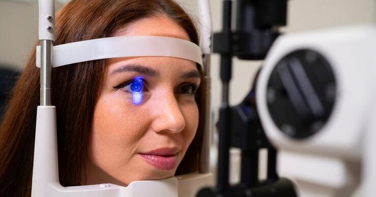
They utilized the PLEX Elite 9000 to conduct swept-source optical coherence tomography (SS-OCT) angiography on all eyes. The study compared macular neural and microvascular alterations in T2DM patients without retinopathy to age-matched controls. They quantitatively analyzed the acquired images, measuring the thickness of each retinal layer and evaluating macular vascular indices within different capillary plexuses.
The results showed that 49 T2DM patients and 51 age-matched controls participated. T2DM patients demonstrated a notable decrease in the mean macular thickness of the ganglion cell-inner plexiform layer (GC-IPL) (82.5 ± 5.5 μm vs. 86.2 ± 5.0 μm, P=0.001) and macular retinal nerve fiber layer (RNFL) (45.8 ± 3.0 μm vs. 48.1 ± 3.7 μm, P=0.001). Moreover, macular full retinal thickness was significantly lower in diabetic eyes compared to controls (324.9 ± 16.3 μm vs. 332.8 ± 13.7 μm, P=0.009). Vascular measurements unveiled subtle alterations in macular vascular skeleton density within the total capillary plexuses in T2DM patients (0.132 ± 0.005 vs. 0.135 ± 0.005, P=0.019).
Investigators concluded that SS-OCT metrics, especially macular RNFL and GC-IPL thickness, showed promise for early DR detection in T2DM without signs, warranting further study.
Source: ajo.com/article/S0002-9394(24)00090-4/abstract