
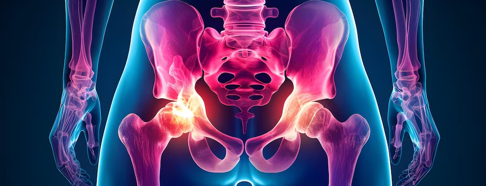
To explore the possibility of using a novel technique, CT perfusion imaging, to monitor the response to anti-tubercular therapy (ATT) in patients with intestinal tuberculosis.
A prospective observational study was performed in adults with treatment naive-intestinal tuberculosis. Clinical, endoscopic, and conventional radiological findings of patients were compared at baseline and post-ATT. CT perfusion imaging was performed with recording of six perfusion parameters (blood flow, blood volume, mean transit time, time to peak, maximum peak intensity, and permeability/blood flow extraction).
Twenty-two patients (13 women, 59%) with a median age of 25 years were recruited. The terminal ileum and ileocaecal junction were the most frequent sites of involvement (59%), with multiple segments of the intestine being involved in 16 patients (73%). Median duration of ATT was 6 months (range 6-10 months). Complete clinical response was observed in 22/22 (100%) patients, endoscopic response in 12/12 (100%) patients, and radiological response in 10/13 (76%) patients. There was a significant decrease in mean blood flow, blood volume, maximum peak intensity, and an increase in mean transit time and time to peak on follow-up CT perfusion imaging performed after 6 months of ATT.
Significant alterations in CT perfusion parameters were demonstrated following treatment, consistent with a decline in inflammation and vascularity. CT perfusion imaging of the bowel is a novel means to assess the radiological response to ATT in intestinal tuberculosis, although at the cost of a higher dose of radiation exposure.
Copyright © 2023. Published by Elsevier Ltd.
Assessment of CT perfusion indices of the clinicoradiological response to anti-tubercular therapy in patients with intestinal tuberculosis.
Oct 16, 2023
REFERENCES & ADDITIONAL READING
PubMed
MORE ARTICLES BELOW
PW Weekly Newsletters
The latest articles and insights from your colleagues in your specialty(ies) of choice.
RECOMMENDED ARTICLES FOR YOU


Advertisement


PW PODCAST
Pre-Nups for Physicians, ACC 2024 Late-Breakers
May 08, 2024What & Why You Need to Know About Value-Based Care
Apr 10, 2024Advertisement


MORE ARTICLES BELOW
PW Weekly Newsletters
The latest articles and insights from your colleagues in your specialty(ies) of choice.
RECOMMENDED ARTICLES FOR YOU
PW PODCAST
Pre-Nups for Physicians, ACC 2024 Late-Breakers
May 08, 2024What & Why You Need to Know About Value-Based Care
Apr 10, 2024Advertisement


Stay connected to the latest news with Physician’s Weekly
Insights from the leaders in medical research, trending topics in clinical medicine, and perspectives from your colleagues.
Subscribe to our free Newsletters to receive weekly emails, and even get a laugh or two from our medical cartoons.